Receptor-Mediated Endocytosis
What is Receptor-Mediated Endocytosis
Receptor-mediated endocytosis (RME), also known as ‘Clathrin-mediated endocytosis’, is a process by which bulk of specific molecules like metabolites, vitamins, hormones, proteins, and also viruses (in some cases) get imported into a cell through binding to a receptor present on the cell surface.
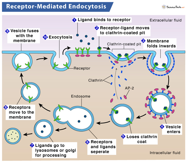
These molecules adhere to receptors and enter the cell through the infolding of the plasma membrane, which eventually gets pinched off into a vesicle. Here an essential structural protein, clathrin, coats the budding vesicle giving it its distinctive spherical form.
Since RME is a form of endocytosis that helps to ship molecules into the cell from the extracellular matrix through a clathrin-coated vesicle, it is alternatively called Clathrin-mediated endocytosis.
Where It is Found
It is a significant activity of the plasma membranes of eukaryotic cells. As the name suggests, the cell surface receptors bind with their respective molecules to internalize them in this process.
Is It a Form of Active or Passive Transport
RME is a form of active transport since it requires energy (ATP) to transport large quantities of macromolecules inside the cell.
How does Receptor-Mediated Endocytosis Work
RME is a type of targeted endocytosis. It involves transmembrane receptor proteins in the plasma membrane that possess a specific binding affinity for certain substances.
These receptors occur in clusters in particular regions of the plasma membrane called coated pits. Clathrin, a protein located on the cytoplasmic side of a cell, coats the pit. Hence, they are known as clathrin-coated pits.
Once the targeted molecule binds to the receptor, the internalization of the pit region begins, and clathrin-coated vesicles form. These vesicles get fused with early endosomes (membrane-bound sacs that help to sort incorporated material). Consequently, the clathrin coating gets removed. As a result, the contents get emptied into the cell.
The uptake of specific compounds is dependent on the receptor involved in the process. If the process fails, the material will not get eliminated from the tissue fluids or blood. Instead, it will stay there and increase in concentration.
Steps
- At its initial stage, a molecule binds to its respective receptor molecule on the cell membrane.
- After binding, a signal triggers a phospholipid component (PIP2- Phosphatidylinositol 4, 5-bisphosphate) to adhere to the adaptor protein (AP2). As a result, the adaptor protein undergoes conformation changes.
- The structural change in adaptor protein allows it binds to the receptors on the cell membrane and recruit clathrin.
- The receptor-bound molecule (ligand) then travels along the membrane to the clathrin-coated pit. Clathrin is a three-legged molecule consisting of three heavy chains and three light chains.
- Upon reaching the pit, the ligand begins to fold inwards as clathrin binds with more adaptor proteins. Consequently, a part of the membrane gets dissociated by the action of a pair of molecular scissors called dynamin (GTPase). The closed coated vesicle thus formed encapsulates the ligand-receptor complex, adaptor proteins, and extracellular fluid.
- Next, the vesicles fuse with the endosome in the cytoplasm, and its clathrin coating gets removed. This step depends on the type of molecule. For example, if the receptor-bound molecule is a pathogen, the opsonization mechanism is activated, leading to its destruction. As the process gets over, the protein coat of clathrin falls off to allow the vesicle to merge with an early endosome.
- As the vesicles get fused with the endosome, multiple compartments start to form within it. Simultaneously, the molecule detaches itself from the receptor.
- Due to a series of chemical changes, the early endosome gets converted into a late endosome. The late endosome then splits into two, one containing the substrate molecule, while the other contains the receptor.
- The one that contains the substrate molecule combines with a lysosome. The hydrolytic enzymes present in the lysosome break down the molecule into simpler components, thus causing cell digestion. The end products of enzyme degradation get released into the cytoplasm, which the cell later uses for various activities.
- Finally, the endosome containing the receptor is recycled and returned to the cell membrane. The receptors are then capable of repeating the process all over again.
Functions and Examples
RME is more than 100-times more efficient for taking in selective molecules than pinocytosis in a cell. Some of its most important functions and examples are given below:
Uptake of LDL and Iron in Mammalian Cells
Cholesterol, being insoluble in body fluids, is transported into the mammalian cell through RME. It is transported in as low-density lipoprotein, the primary plasma carrier of cholesterol in the body.
After incorporation, the LDLs get transported to lysosomes, degraded into amino acids, forming cholesterol and fatty acids. Finally, the cholesterol thus formed is directly incorporated into cell membranes or is reesterified and stored as lipid droplets in the cell for future use.
RME also helps in the uptake of iron through the endocytosis of transferrin (an iron-binding protein) at the cell surface, with the help of the transferrin receptor (TfR). The process of uptake is similar to LDL.
Uptake of Large Biomolecules and Growth Factors
Several biomolecules are imported into the cell by RME. For example, glucose, the body’s primary energy source, is transported by the glucose transporter-1 receptor. An l-type amino acid transporter-1 receptor transports amino acids. Specific receptors also transport thiamin, biotin, folic acid, vitamin B12, and neuropeptides. Insulin-like growth factors, such as IGF-I, IGF-II, and leptin, are transported by the endogenous receptors located in the brain vascular endothelium.
Regulating Cell Signaling
RME of signaling receptors is a crucial mechanism to regulate signaling in cells. It can attenuate the strength or duration of many plasma membrane-regulated signaling processes by physically reducing the concentration of cell surface receptors accessible to the ligand. However, in some cases, it shifts the dose-response relationship. As a result, a higher concentration of a ligand is required to trigger a response of the same magnitude.
Facilitates Entry of Certain Viruses and Other Foreign Antigens
Several pathogens like bacteria and viruses generally enter the host cell for replication by exploiting the endocytosis machinery. However, unlike bacteria, viruses use the cell’s signaling mechanisms and regulatory pathways to induce endocytosis and prepare the host cell for invasion.
FAQs
Ans. In pinocytosis, vesicles form at the plasma membrane and bring fluids and small molecules into the cell. Thus the process is known as cell drinking. In phagocytosis, vesicles form at the plasma membrane to bring solid particles into the cell. Thus the process is often called cell eating. Finally, in receptor-mediated endocytosis, specific target molecules bind to cell surface receptors proteins called clathrin, form vesicles that subsequently help to internalize the cells. Thus the process is also called Clathrin-mediated endocytosis.
-
References
Article was last reviewed on Friday, February 17, 2023


