Parts of a Neuron and Their Function
Neurons or nerve cells are the basic building blocks or units of the nervous system. Nearly 86 billion neurons work co-ordinately within the nervous system to keep the body organized. They are highly specialized cells that act as information processing and transmitting units of the brain. A group of neurons forms a nerve.
What are the Different Parts of a Neuron

Although they have a characteristic elongated shape, they vary widely in size and properties based on their location and type of functions they perform.
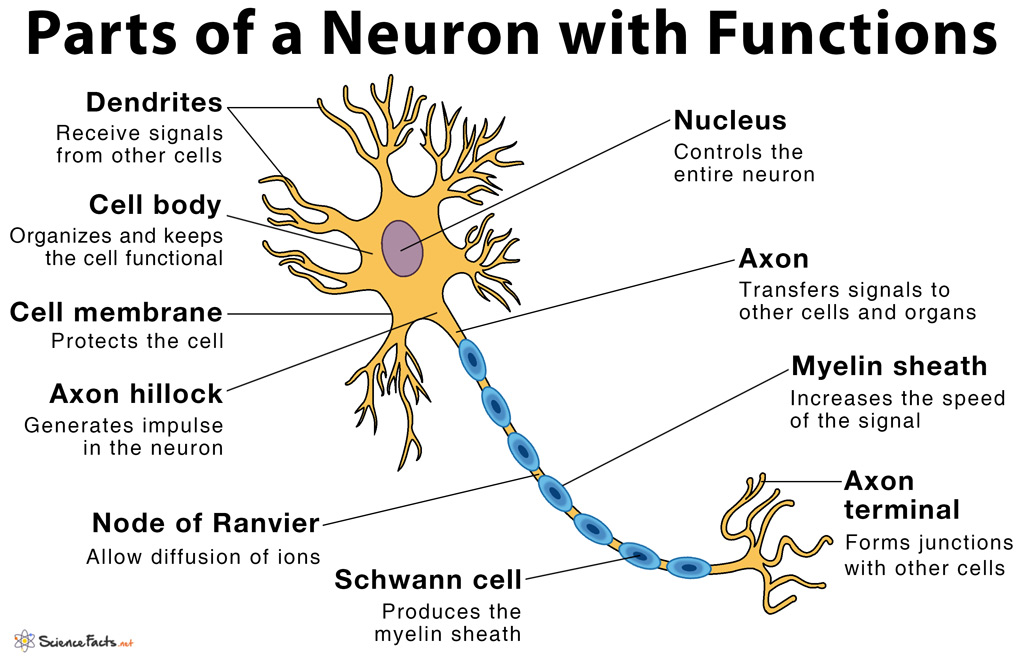
While they have the common features of a typical cell, they are structurally and functionally unique from other cells in many ways. All neurons have three main parts: 1) dendrites, 2) cell body or soma, and 3) axons. Besides the three major parts, there is the presence of axon terminal and synapse at the end of the neuron.
1) Dendrites
They are specialized extensions that resemble the branch of a tree. Dendrites help to increase the surface area available for connections with the adjacent neurons and thus in receiving incoming signals from them. Some neurons have very small and short dendrites, while in others, they are very long. Most neurons have numerous dendrites, while a few others have only one of them.
Functions
- Acquiring chemical impulse from other cells and neurons
- Converting the chemical signals into electrical impulses
- Carrying electrical impulses towards the next part of the neuron, the cell body
2) Cell Body or Soma
It is the core of the neuron, similar to a cell that contains the nucleus and all other cellular organelles. The cell body is also the largest part of a neuron enclosed by a cell membrane that protects the cell from its immediate surroundings and allows its interaction with the outside environment. They attach to all the dendrites and thus integrate all the signals. All metabolic activities of the cell take place in the cell body, which also contains DNA, the genetic material of the neuron.
Functions
- Supporting and organizing the functions of the whole neuron
- Joining the signals received by the dendrites and passing them to the axons, the next part of the neuron
3) Axons
They are fine, elongated fiber-like extensions of the nerve cell membrane. Axons run from the cell body of one neuron until the terminal of the next neuron. They are the longest part of the neuron that varies widely in length, with some are as tiny as 0.1 millimeters, while some others can be over 3 feet. The larger the diameter of the axon, the faster is the rate of transmission of nerve signals.
Sometimes, a single axon is highly branched to allow better communication with multiple target neurons at the same time.
Parts of an Axon
a) Axon hillock – The part of the axon which remains attached to the cell body or soma.
b) Myelin sheath – The layer of fatty acid produced from specialized cells called Schwann cells that are wrapped around the axon.
c) Nodes of Ranvier – The gaps between the discontinuous myelin sheath that is running along the axon.
Functions
- Axons help to receive signals from other neurons and transmit the outflow of the message to the adjacent connected neurons and also to other muscles and glands by changing the electrical potential of the cell membrane called the action potential.
- Myelin sheath insulates the axon and thus prevents shock similar to an insulated electric wire
- Myelin sheath also increases the speed of the flow of signals through the axon
- Nodes of Ranvier allows diffusion of ions in and out of the neuron and thus maintains the electrical potential of the neuron
Apart from all the major parts described so far in a neuron, there are other structures (that are not basic parts) found at the junctions between the neurons that help to establish functional links or connections between them. They are described below.
Connecting Parts of a Neuron: Axon Terminal and Synapse
Axon terminal also called synaptic bouton or terminal button is the terminal branches of the axon located at the very end of the neuron. They are farthest from the soma and contain chemical messengers called neurotransmitters in specialized structures called synaptic vesicles.
Synapse or synaptic cleft is the small space or gap between two adjacent neurons. It is formed between the axon terminal of one neuron and the dendrites of the next neurons.
Functions
- Releasing of neurotransmitters through specific transport vesicles, called synaptic vesicles from one neuron to the adjacent connected neurons called exocytosis
- Synaptic vesicles of one neuron for conducting nerve impulse to the adjacent connected neuron through exocytosis
- Sending neuronal information from one nerve cell to another and also to other cells of the muscle or gland with the help of neurotransmitters
- Re-up taking of excessive neurotransmitters from the synaptic cleft
-
References
Article was last reviewed on Thursday, February 2, 2023


What is the function of each part ?
Check the post to know more
What is the name of the nerve cell
It is also called neuron.
The lesson gives detail and vital information about neuron and its structures. My question is what inside axon. Is that nerve fiber. Thank you.
Best
Yes. Axons are thin nerve fibers that connects neurons so that they can communicate.
This is so helpful! Thank you so much!🐈⬛
very intresting and important information